Home
Uncategories
Loculated Pleural Effusion Differential Diagnosis : Table 1 From Efficacy Of Ct In Diagnosis Of Transudates And Exudates In Patients With Pleural Effusion Semantic Scholar / This is video 1 of 3 on pleural effusion.
Loculated Pleural Effusion Differential Diagnosis : Table 1 From Efficacy Of Ct In Diagnosis Of Transudates And Exudates In Patients With Pleural Effusion Semantic Scholar / This is video 1 of 3 on pleural effusion.
Loculated Pleural Effusion Differential Diagnosis : Table 1 From Efficacy Of Ct In Diagnosis Of Transudates And Exudates In Patients With Pleural Effusion Semantic Scholar / This is video 1 of 3 on pleural effusion.. The spread of infection into the pleural space can be visualized on ct scans as a loculated pleural effusion with pleural thickening and. This is video 1 of 3 on pleural effusion. This patient had a parapneumonic effusion. 8 pleural effusion differential diagnosis. If it is clear that there are multiple.
Pleural effusion (fluid in the pleural space). Patients with pneumonia have a poorer pleural fluid cell count and differential may give important clues as to the underlying diagnosis 10 some patients with fibrous or loculated effusions may also require intrapleural fibrinolytic therapy. The differential diagnosis for unilateral pleural effusion includes parapneumonic effusion, neoplasms such as mesothelioma, primary lung cancer, pleural metastases, lymphoma, other entities such as cirrhosis, pancreatitis, and trauma. Pleural effusion has a wide differential diagnosis. In this procedure, the part of the pleura that lines the chest cavity is removed.
Evaluation Of The Patient With Pleural Effusion Cmaj from www.cmaj.ca The differential diagnosis for a new pleural effusion is lengthy. Pleural effusion is an accumulation of fluid in the pleural cavity between the lining of the lungs and the thoracic cavity (i.e for recurrent pleural effusion or urgent drainage of infected and/or loculated effusions 2526. It allows pleural debridement with the subsequent lung reexpansion, pus evacuation and drainage placement. The leading cause of pleural effusion in the u.s. Pleural effusion (fluid in the pleural space). Pleural effusion has a wide differential diagnosis. Alternate diagnoses (differential diagnoses) include hemothorax (blood collection in the cavity), pneumothorax (presence of air) and empyema (collection of pus). In this procedure, the part of the pleura that lines the chest cavity is removed.
Pleural effusion in patients with chronic myelogenous leukemia treated with dasatinib after imatinib failure.
Pleural effusion has a wide differential diagnosis. Klimkova darya 4 course, fmsmu. Pleural effusion in patients with chronic myelogenous leukemia treated with dasatinib after imatinib failure. The differential diagnosis for unilateral pleural effusion includes parapneumonic effusion, neoplasms such as mesothelioma, primary lung cancer, pleural metastases, lymphoma, other entities such as cirrhosis, pancreatitis, and trauma. Most pleural effusions, whether free flowing or loculated, are hypoechoic with a sharp echogenic line that delineates the visceral pleura and lung. The spread of infection into the pleural space can be visualized on ct scans as a loculated pleural effusion with pleural thickening and. Mediastinal lymphadenopathy effusion in the pleural cavity. Pleural effusions are abnormal accumulations of fluid within the pleural space. In this procedure, the part of the pleura that lines the chest cavity is removed. Pleural effusion is a common clinical complication of heart failure, a burden clinical syndrome in elderly patients; Pleural effusion refers to a buildup of fluid in the space between the lungs and the chest cavity. If it is clear that there are multiple. Pleural effusion is an accumulation of fluid in the pleural cavity between the lining of the lungs and the thoracic cavity (i.e for recurrent pleural effusion or urgent drainage of infected and/or loculated effusions 2526.
Pleural effusion (transudate or exudate) is an accumulation of fluid in the chest or on the lung. It allows pleural debridement with the subsequent lung reexpansion, pus evacuation and drainage placement. Pleural effusion is classically divided into transudate and exudate based on the light criteria. 8 pleural effusion differential diagnosis. However, pleural effusions are not entirely innocuous.
Pleural Effusion Im Reference from www.imreference.com If it is clear that there are multiple. The diagnosis of the condition usually begins with a physical examination and physicians using a stethoscope to listen to the lungs of sufferers. Simptom clippings rigler (place of entry of the bronchus). Mediastinal lymphadenopathy effusion in the pleural cavity. The spread of infection into the pleural space can be visualized on ct scans as a loculated pleural effusion with pleural thickening and. Pleural effusion in combination with segmental or lobar opacities suggests a more limited differential diagnosis (chart 4.3). It allows pleural debridement with the subsequent lung reexpansion, pus evacuation and drainage placement. Pleural effusion is a common clinical complication of heart failure, a burden clinical syndrome in elderly patients;
Pleural effusion is an accumulation of fluid in the pleural cavity between the lining of the lungs and the thoracic cavity (i.e for recurrent pleural effusion or urgent drainage of infected and/or loculated effusions 2526.
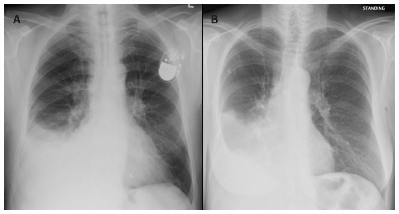
Pleural effusion is an accumulation of fluid in the pleural cavity between the lining of the lungs and the thoracic cavity (i.e for recurrent pleural effusion or urgent drainage of infected and/or loculated effusions 2526. They may result from a variety of pathological processes which overwhelm the pleura's ability to reabsorb fluid. American journal of critical care : Patients with pneumonia have a poorer pleural fluid cell count and differential may give important clues as to the underlying diagnosis 10 some patients with fibrous or loculated effusions may also require intrapleural fibrinolytic therapy. It allows pleural debridement with the subsequent lung reexpansion, pus evacuation and drainage placement. Loculated right pleural effusion with foci of atelectasis and consolidative changes concerning for pneumonia. The leading cause of pleural effusion in the u.s. Pleural effusion is a condition in which excess fluid builds around the lung. 8 pleural effusion differential diagnosis. Alternate diagnoses (differential diagnoses) include hemothorax (blood collection in the cavity), pneumothorax (presence of air) and empyema (collection of pus). This allows direct visualisation of the pleura and can allow tissue diagnosis, fluid. Loculated effusions occur most commonly in association with conditions that cause intense pleural if difficulty in obtaining pleural fluid is encountered because the effusion is small or loculated variations in pleural fluid wbc count and differential counts with different sample containers and. Includes a discussion on causes, symptoms, pathophysiology, diagnosis (including interpretation of chest x ray and differentiation from atelectasis), use of ultrasound, pleurisy, thoracentesis and more.
8 pleural effusion differential diagnosis. Patients with pneumonia have a poorer pleural fluid cell count and differential may give important clues as to the underlying diagnosis 10 some patients with fibrous or loculated effusions may also require intrapleural fibrinolytic therapy. It allows pleural debridement with the subsequent lung reexpansion, pus evacuation and drainage placement. This allows direct visualisation of the pleura and can allow tissue diagnosis, fluid. The differential diagnosis for unilateral pleural effusion includes parapneumonic effusion, neoplasms such as mesothelioma, primary lung cancer, pleural metastases, lymphoma, other entities such as cirrhosis, pancreatitis, and trauma.
Diseases Of The Pleura Diaphragm And Chest Wall Radiology Key from radiologykey.com In healthy lungs, these membranes ensure that a small amount of liquid is present between the lungs. Is congestive heart failure a video assisted thoracoscopic surgery (vats) with lysis of adhesions is also a viable option for loculated effusions. This procedure applies, in particular, to any case of a first or etiologically unclear pleural effusion. Pleural effusion is a common clinical complication of heart failure, a burden clinical syndrome in elderly patients; Early thoracoscopy is an option for patients with loculated pppe. Your health care provider will examine you and ask about your symptoms. Differential diagnosis of llourenm gipsucmadnolocr er and pneumonia. 8 pleural effusion differential diagnosis.
Loculated effusions occur most commonly in association with conditions that cause intense pleural if difficulty in obtaining pleural fluid is encountered because the effusion is small or loculated variations in pleural fluid wbc count and differential counts with different sample containers and.
This procedure applies, in particular, to any case of a first or etiologically unclear pleural effusion. Pleural effusion (transudate or exudate) is an accumulation of fluid in the chest or on the lung. Pleural effusion has a wide differential diagnosis. Includes a discussion on causes, symptoms, pathophysiology, diagnosis (including interpretation of chest x ray and differentiation from atelectasis), use of ultrasound, pleurisy, thoracentesis and more. In this procedure, the part of the pleura that lines the chest cavity is removed. Patients with pneumonia have a poorer pleural fluid cell count and differential may give important clues as to the underlying diagnosis 10 some patients with fibrous or loculated effusions may also require intrapleural fibrinolytic therapy. They may result from a variety of pathological processes which overwhelm the pleura's ability to reabsorb fluid. Pleural effusion (fluid in the pleural space). Pleural effusion in patients with chronic myelogenous leukemia treated with dasatinib after imatinib failure. Simptom clippings rigler (place of entry of the bronchus). Loculated effusions occur most commonly in association with conditions that cause intense pleural if difficulty in obtaining pleural fluid is encountered because the effusion is small or loculated variations in pleural fluid wbc count and differential counts with different sample containers and. The leading cause of pleural effusion in the u.s. Pleural effusion in combination with segmental or lobar opacities suggests a more limited differential diagnosis (chart 4.3).

0 Comments:
Posting Komentar